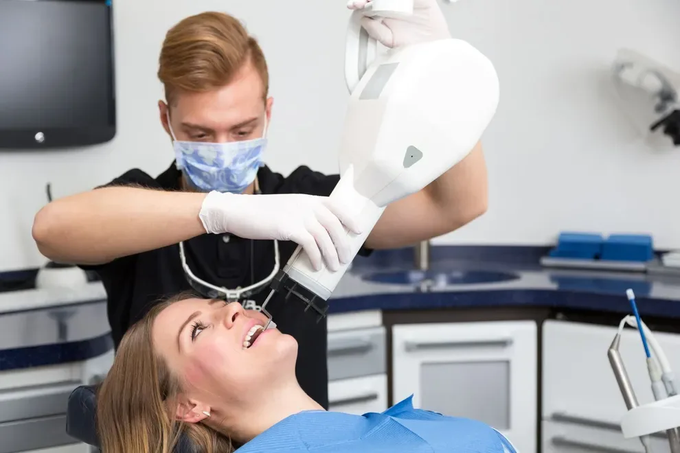
The Latest in Dental Imaging - Digital Dental Radiography

Table of Contents
- How Does It Work?
- Types
- Uses
- How Often Do You Need It?
- Benefits
- Disadvantages
- Training Required
- References
Digital dental radiography is a process that helps produce images of your mouth, just like a regular X-ray. In addition, it scans your mouth almost instantly and stores the digital images on a computer.
The process uses sensors connected to a computer to generate clearer gray-scale images. It makes it easier for the dentist to monitor, detect, diagnose, and treat oral diseases and conditions.
How Does It Work?
Unlike traditional X-rays that requires film to produce the image, digital dental radiography uses digital X-ray sensors to create enhanced images. It combines electronic sensors and bursts of radiation that pass through soft tissue but are reflected by bones, making them visible. There are three main methods of achieving this:
Direct: records images through a digital sensor placed in the mouth.
Indirect: a scanner surveys traditional dental X-rays and converts them into digital format.
Semi-indirect: combines both digital sensor and scanner to convert an X-ray into digital images.
Although it uses bursts of radiation, the typical bitewing dental radiography exposure is so minor it doesn’t exceed 1/3000 times the exposure you get through natural means.
Types
There are two main types of digital dental radiographs–-those taken inside the mouth and those taken outside.
Of the two, intraoral is the most common type of dental X-rays. They help check the status of developing teeth, detect cavities, and monitor bone and tooth health.
Extraoral X-rays help identify potential problems to the bone, teeth, and jaw, temporomandibular joints, and help monitor jaw development. Types of intraoral X-rays include:
Bitewing X-rays. This involves a patient biting down on film, and the results show details of the upper and lower teeth. A bitewing X-ray can show a tooth’s details from the crown to the supporting bone.
Periapical (limited) X-rays. These show a tooth from its crown to the root and surrounding bone. This type of X-ray helps detect abscesses and bone loss.
Types of extraoral include:
Panoramic X-rays. These involve a machine rotating around the head and showing the whole upper and lower dental region in one image. These X-rays help dentists plan for dental implants, detect jaw problems and impacted teeth.
Multi-slice computed tomography (MCT). This X-ray shows a “slice” of the mouth while blurring other parts. This X-ray helps examine parts of the mouth that are otherwise difficult to see.
Uses-Why Would I Need Digital X-rays?
During a dental exam, a digital X-ray allows your dentist to look at the condition of your mouth to detect potential problems or monitor your oral health.
A digital X-ray provides accurate, real-time results that allow for a quicker diagnosis. Traditional X-rays require a waiting period that may often require a second dental appointment.
How Often Do You Need to Conduct Digital Dental Imaging?
It depends on several factors such as your general oral health, medical and dental history. If you have chronic dental issues such as abscess, tooth decay, and uneven teeth, dentists recommend an X-ray every six months.
For a healthier individual experiencing no dental health issues, dentists suggest an X-ray every two-to-three years. New patients require a dental X-ray on their first visit, while others only need an X-ray as needed.
Benefits of Digital Dental Radiographs
Compared to traditional X-rays, digital dental radiographs have the following benefits:
You can instantly view results from a dental radiograph on a screen, enhance the image detail, and transmit them electronically without losing quality. Dental technicians can also increase the image size without altering information.
Digital radiography offers a more streamlined view of the oral structure as it covers more angles. It allows dentists to detect problems earlier on, saving time and money. Digital X-rays detect issues related to previous dental work, something that a traditional X-ray cannot do without multiple dental visits.
Digital storage technology allows for more extensive data storage capacity on small drives. You can send digitized data to another office instantly for further reference.
Digital X-rays do not use any chemical processing, unlike traditional X-rays, offering an eco-friendlier and greener alternative.
Digital sensors are more sensitive to X-radiation and require up to 80% less radiation than traditional X-rays.
Shorter appointment times because no preparation or waiting is required.
Improved diagnostics resulting from clearer images enhance the quality of treatment and success rate.
Disadvantages
Setting up digital dental radiography is expensive for a practice, costing up to $15,000 for a wired sensor system and up to $50,000 for a wireless one. This price excludes the cost of software and other hardware such as computers, servers, and repairs and maintenance. A dental practice often has more than one sensor, meaning the cost goes higher. Damaged and worn sensors are also costly to replace.
As digital dental radiography is still relatively new, it requires additional training that needs constant refreshing to keep up with current technology.
Digital sensors are sensitive to scattered radiation, affecting the images.
Digital dentist radiography is a recent technology and is not universally applicable across all dental clinics.
Photostimulable phosphor (PSP) plates are thinner than film plates used for traditional X-rays. The thinner plates are fragile, wear out quicker, and require frequent replacing.
Some sensors are bulkier than dental films, leading to discomfort for patients, especially those with problematic gag reflexes.
It is not easy to sterilize PSP plates and digital sensors, and they require a protective barrier for every use. Dentists must practice caution to prevent infection and cross-contamination.
Lack of availability of post-processing functions.
Training Required
Dentists, dental assistants, oral radiologists, and dental hygienists all need training before performing digital radiographs. However, the training is less stringent than that required of medical X-ray, and personnel can access this training from dental boards and universities.
The course requires that the practitioner be at least 18 years old and have at least three months hands-on assistance training under a licensed dentist. Personnel will need to fill in a dental qualification form before starting the course, which is three-to-seven hours.
Topics in the training include:
Historical background of X-rays
Digital dental terminology
Digital X-ray machine components
Digital X-ray shooting techniques
Interpreting digital dental X-rays
Sensor and equipment placement
Digital image processing and storage
Digital presentation and display
Infection control
Upcoming trends in digital dental radiography
